


- Diagnostics
Accurate diagnosis for your eyes
- Diagnostics
Accurate diagnosis for eye health
Specialised eye examinations to assess vision, detect diseases early and monitor eye health. Every examination helps determine the right next steps for your care.
- Diagnostics
Every examination performed with care and attention
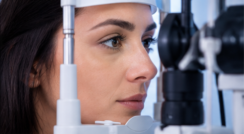
Visual Acuity Testing
Visual acuity testing is one of the basic steps in an eye examination. Appropriate tests assess near and distance vision, helping identify difficulties that may require optical correction or further examination.
Conditions that may be assessed:
- Myopia (short-sightedness))
- Hyperopia (long-sightedness)
- Astigmatism
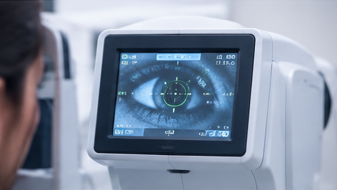
Computerised Refraction Measurement
Computerised refraction uses a specialised device to assess how light is focused by the eye. The results help identify possible refractive errors and guide further examination. Additional tests may be performed to determine the precise optical correction required.
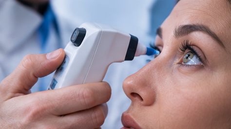
Intraocular Pressure (IOP) Measurement
Intraocular pressure is the pressure of the fluid inside the eye. Measuring it is an important part of glaucoma assessment, as glaucoma can damage the optic nerve and cause vision loss. The result is interpreted alongside other examinations, since a single measurement cannot confirm or rule out glaucoma.
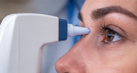
Tear Production Measurement
Tears lubricate, protect and maintain the surface of the eye. Measuring tear quantity can help assess insufficient tear production, which may be associated with dryness, burning, irritation or a foreign-body sensation. The results help guide appropriate care.
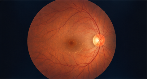
Direct fundoscopy
Direct fundoscopy allows the internal structures of the eye to be examined, particularly the optic nerve and retinal blood vessels. It can help identify changes associated with various eye diseases or general health conditions.
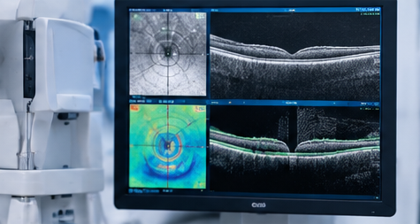
OCT (Optical Coherence Tomography)
OCT is a non-invasive examination that uses light waves to create detailed images of the layers of the eye. Depending on the equipment and examination protocol, it can assess the macula, optic nerve, cornea and anterior chamber angle.
OCT helps detect and monitor structural changes associated with various eye conditions.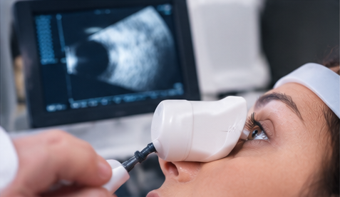
Ocular Ultrasound Examination
Ocular ultrasound uses sound waves to provide information about the internal structures of the eye. Depending on the type of examination, it may be used to assess eye anatomy, measure the length of the eye or determine corneal thickness.
Types of examinations:- A-scan – Used to measure the length of the eye.
- B-scan – Produces images of the internal structures of the eye.
- Pachymetry – Measures corneal thickness.
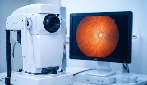
Fundus Photography
Fundus photography captures images of internal eye structures, including the retina, blood vessels and optic disc. These images can document the baseline condition and help compare changes during follow-up examinations.
Detailed Fundus Examination
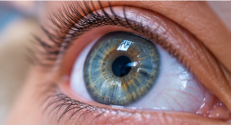
A detailed fundus examination helps assess the retina, blood vessels and optic nerve. It is particularly important for patients with chronic diseases or conditions that may affect eye health.
Particular attention is paid to changes that may be associated with:
- Diabetes and diabetic retinopathy
- Hypertension and vascular changes
- Autoimmune diseases
- Intraocular injuries
- Other retinal changes
- Ocular procedures
Contact Lens Fitting
- Types of lenses:
- Soft contact lenses
- Soft toric contact lenses
- Rigid GP contact lenses
- Toric GP contact lenses
- GP contact lenses for keratoconus

Interested in our services? Contact us today!
Pediatric Ophthalmology
- © 2026 Ordinanca Vizioni
- Web design Primeframe